

Cranial Fossa Anatomy

Q. Posterior Cranial fossa Boundaries ?
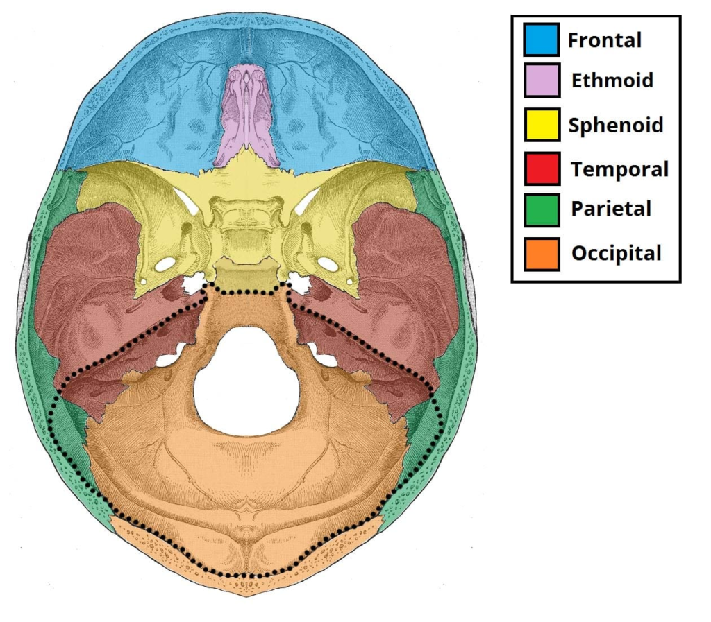
The boundaries of the posterior cranial fossa are:
Anterior:
– Clivus: The sloping anterior wall of the posterior fossa formed by the basilar occiput and the dorsum sellae of the sphenoid.
– Petrous temporal bone: The dense, pyramid-shaped portion of the temporal bone that contains the inner ear structures.
Posterior: Occipital bone: The bone that forms the back and base of the skull.
Lateral:
– Squamous temporal bone: The upper, flat portion of the temporal bone.
– Mastoid temporal bone: The irregular, protruding portion of the temporal bone behind the external ear.
Superior: Tentorium cerebelli: A fold of dura mater that separates the cerebellum from the occipital lobes of the brain.
Inferior: Foramen magnum: The large opening in the occipital bone that allows the spinal cord to connect with the brain.
The posterior cranial fossa contains the cerebellum, pons and medulla oblongata of the brainstem. It is the deepest fossa (dent) within the skull.
The clivus and petrous temporal bones form its sloping anterior wall. The occipital bone forms the posterior wall and also contributes to the foramen magnum inferiorly with the lateral parts of the exoccipitals.
The squamous and mastoid temporal bones form the lateral walls. Superiorly, the posterior fossa communicates with the supratentorial compartment through the tentorial notch.
The foramen magnum, along with the hypoglossal canal, jugular foramen and foramen lacerum, provide passages for the exit and entry of important neural and vascular structures related to the posterior fossa.
Q. Bones of posterior cranial fossa ?
The bones that form the posterior cranial fossa are:
1. Temporal bones: The temporal bones form the lateral walls and parts of the anterior wall of the posterior fossa. The parts of the temporal bone in the posterior fossa include:
– Petrous temporal bone: Forms the anterior wall, contains the inner ear.
– Mastoid temporal bone: Irregular protruding region behind the external ear.
– Squamous temporal bone: Upper, flat portion that forms the lateral wall.
2. Occipital bone: The occipital bone forms the posterior wall and parts of the inferior wall of the posterior fossa. The parts of the occipital bone include:
– Squamous occipital: Forms the posterior wall. Has an external occipital protuberance and groove for sinuses.
– Lateral parts of exoccipitals: Form inferior parts of the walls and contribute to the foramen magnum.
– Basilar occipital: Sloping anterior portion that forms the clivus along with the dorsum sellae.
3. Parietal bones: The parietal bones form a small superior portion of the lateral walls of the posterior fossa where the tentorium cerebelli attaches. They do not directly contribute to the posterior fossa but provide attachment for the tentorium which forms its superior boundary.
In summary, the three bones that significantly contribute to the posterior cranial fossa are the temporal bones (lateral and anterior walls), occipital bone (posterior and inferior walls) and parietal bones (indirectly as an attachment for the tentorium). The occipital bone forms the majority of the posterior fossa compartment.
Q.Venous sinus tracts in posterior cranial fossa?
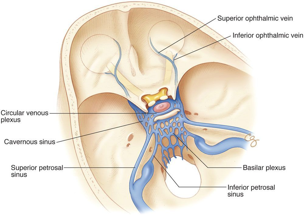
1. Sigmoid sinuses: Paired sinuses that run along the inner surface of the mastoid process of the temporal bone. They exit the posterior fossa through the jugular foramen to continue as the internal jugular veins. The sigmoid sinuses receive blood from the cerebellum, posterior fossa structures and inner ear.
2. Superior petrosal sinuses: Paired sinuses located in the petrous ridge of the temporal bone. They connect the cavernous sinus anteriorly to the sigmoid sinus posteriorly. The superior petrosal sinuses drain the trigeminal ganglion and nerve.
3. Inferior petrosal sinuses: Paired sinuses located between the petrous ridge and basilar occiput. They connect the cavernous sinus to the internal jugular veins. The inferior petrosal sinuses drain parts of the cranial nerves and middle ear.
4. Foramen lacerum and jugular foramen: These are technically foramina, but provide passages for the petrosal sinuses to exit the skull along with CN IX, X and XI.
5. Posterior fossa bridging veins: Small veins that drain the cerebellum and drain either into the petrosal sinuses or directly into the sigmoid/jugular system.
6. Basilar sinus: Midline sinus that runs along the clivus in the midline and drains into the cavernous sinus anteriorly and inferior petrosal/jugular system posteriorly. The basilar sinus receives cerebellar bridging veins along its length.
In summary, the major venous sinus tracts in the posterior fossa include the sigmoid sinuses, superior & inferior petrosal sinuses, basilar sinus and the posterior fossa bridging veins. They drain the cerebellum, brainstem, and cranial nerves and empty into the jugular venous system which exits the skull through the jugular foramen. The petrosal and basilar sinuses also provide connections between the cavernous sinus and jugular system.
Q.foramina and canals in the skull base?
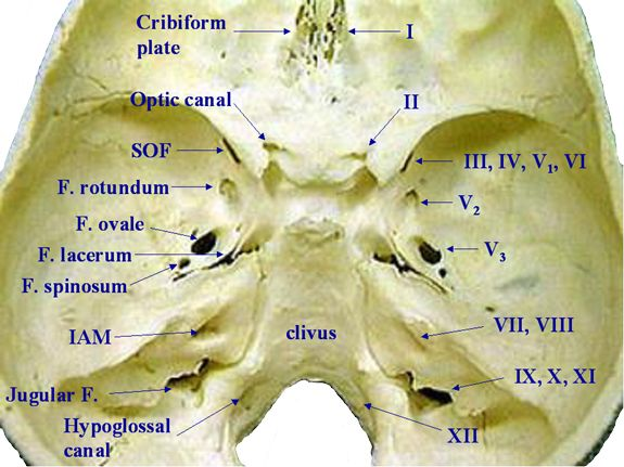
Foramen lacerum:
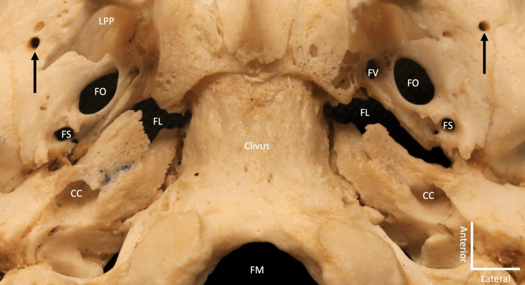
– Inferior part: Filled with fibrocartilage, no structures pass through.
– Superior part: Transmits internal carotid artery, greater and deep petrosal nerves before entering pterygoid canal.
Foramen rotundum: Transmits maxillary division of trigeminal nerve (CN V2).
Foramen ovale: Transmits mandibular division of trigeminal nerve (CN V3), accessory meningeal artery, lesser superficial petrosal nerve, emissary veins.
Foramen spinosum: Transmits middle meningeal artery.
Optic canal: Transmits optic nerve (CN II) and ophthalmic artery. Associated with meningitis causing papilledema.
Superior orbital fissure: Transmits CN III, IV, V1 (nasociliary, frontal and lacrimal nerves), VI and sympathetic nerves.
Stylomastoid foramen: Transmits facial nerve (CN VII) and stylomastoid artery.
Foramen magnum: Transmits vertebral arteries, spinal root of accessory nerve (CN XI), tectorial membrane, anterior and posterior spinal arteries, lower medulla oblongata, apical ligament of the odontoid process.
Jugular foramen: Transmits glossopharyngeal (CN IX), vagus (CN X) and spinal accessory (CN XI) nerves, inferior petrosal sinus and internal jugular vein.
Q : Middle cranial fossa- Contents and Boundaries
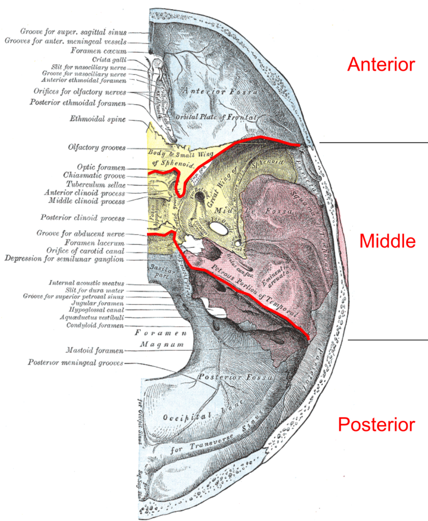
The boundaries of the middle cranial fossa are:
Anterior:
– Lesser wing of sphenoid
– Anterior clinoid processes
Posterior:
– Petrous temporal bone
– Dorsum sellae
Lateral: Squamous part of temporal bone
The contents of the middle cranial fossa include:
– Temporal lobes of the cerebral hemispheres
– Cranial nerves III-VI: As they traverse the lateral wall.
– Internal carotid arteries: Ascend through the carotid canals and emerge to enter the cranium.
– Pituitary gland: Housed in the sella turcica, connected to the hypothalamus by the infundibulum.
– Cavernous sinuses: Venous sinuses located on either side of the pituitary gland. Receive blood from middle fossa structures and contain CN III, IV, V1 and VI.
Foramina transmitting structures:
– Optic canal: Optic nerve (CN II) and ophthalmic artery.
– Superior orbital fissure: CN III, IV, V1, VI and sympathetic nerves.
– Foramen rotundum: Maxillary division CN V (V2).
– Foramen ovale: Mandibular division CN V (V3).
– Foramen spinosum: Middle meningeal vessels.
– Foramina of cribriform plate: Filaments of olfactory nerves (CN I).
The middle cranial fossa forms a major division of the anterior cranial fossa and contains vital neurovascular structures and connections between the brain and orbits/upper face.
Q. ID : Trigiminal impression, Squamous part of the temporal bone ,Tegmen tympani, Frontal crest,Clivus,Groove for the transverse sinus, Internal occipital protuberance, Hypoglossal canal
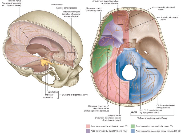
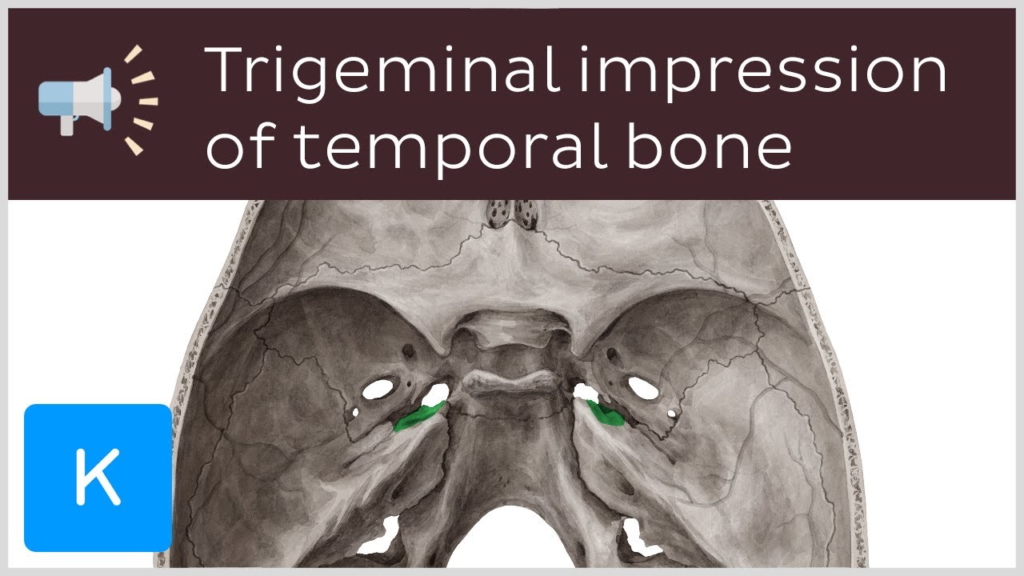
1. Trigeminal impression: Depression on the inner surface of the petrous temporal bone that houses the trigeminal ganglion.
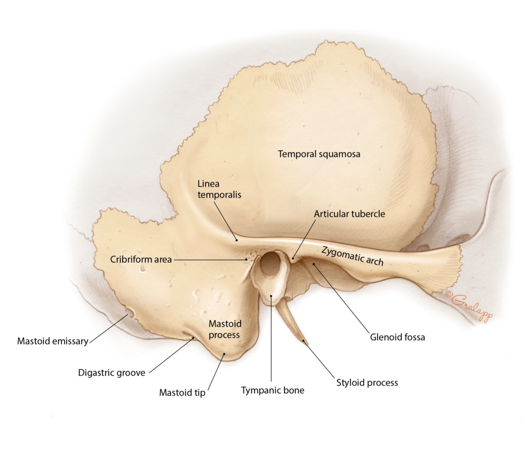
2. Squamous part of temporal bone:
3. Tegmen tympani:
Upper, flat portion of the temporal bone. Forms the lateral wall of the middle cranial fossa.
3. Tegmen tympani:
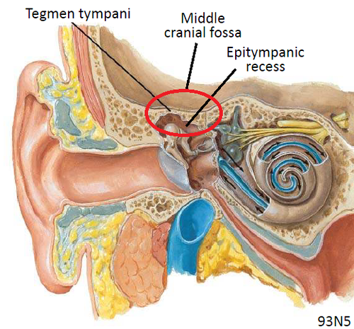
Thin plate of bone that forms the roof of the middle ear cavity. Separates the middle ear from the middle cranial fossa.
4. Frontal crest:
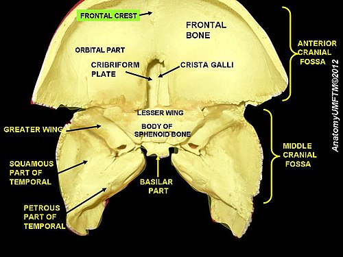
Midline bony ridge on inner surface of frontal bone that forms the attachment for the falx cerebri.
5. Clivus:
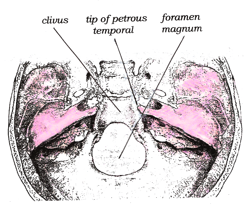
Anterior sloping wall of the posterior cranial fossa formed by the basiocciput and dorsum sellae.
6. Groove for transverse sinus:
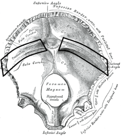
Groove located in the occipital bone that provides passage for the transverse sinuses. The transverse sinuses run laterally in this groove to the sigmoid sinuses.
7. Internal occipital protuberance:
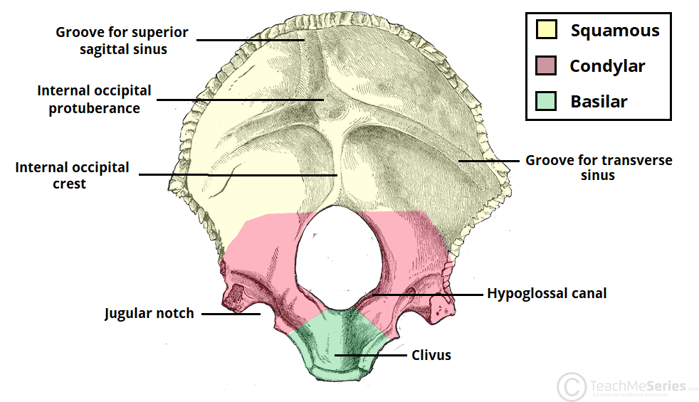
Prominence in the midline on the inner surface of occipital bone. Provides attachment for the falx cerebelli.
8. Hypoglossal canal:
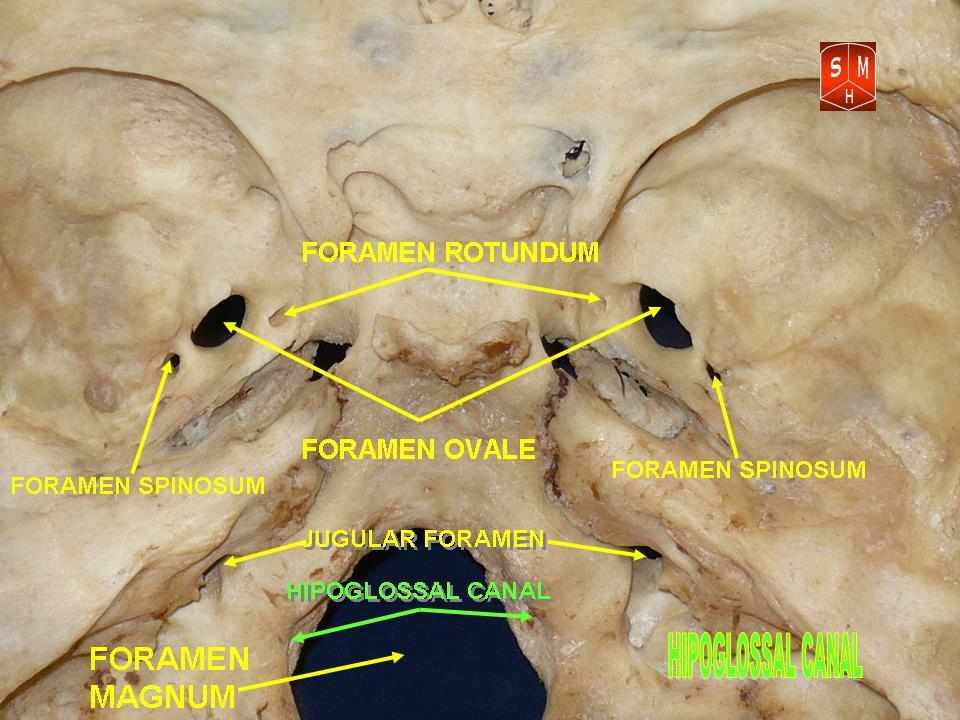
Canal located in the occipital bone that transmits the hypoglossal nerve (CN XII) from the skull to the neck.
In summary:
– The trigeminal impression houses the trigeminal ganglion.
– The squamous part of the temporal bone forms the lateral wall of the middle cranial fossa.
– The tegmen tympani forms the roof of the middle ear.
– The frontal crest provides attachment for the falx cerebri.
– The clivus forms the anterior wall of the posterior cranial fossa.
– The groove for the transverse sinus provides passage for the transverse sinuses.
– The internal occipital protuberance attaches the falx cerebelli.
– The hypoglossal canal transmits the hypoglossal nerve (CN XII) from the skull.
Q.Styloid attachments ?
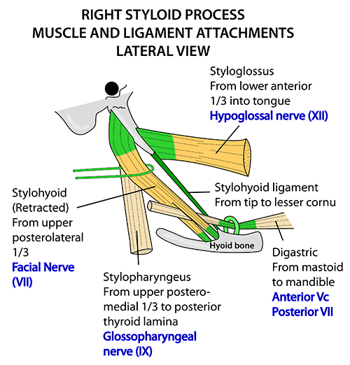
The styloid process of the temporal bone provides attachment for four structures:
1. Styloglossus muscle: A strap muscle that retracts and elevates the tongue. It arises from the styloid process and inserts into the tongue. It is innervated by the hypoglossal nerve (CN XII).
2. Stylohyoid muscle: A small muscle associated with swallowing. It arises from the styloid process and inserts into the hyoid bone. It is innervated by the facial nerve (CN VII).
3. Stylopharyngeus muscle: A muscle located in the lateral pharyngeal wall. It arises from the styloid process and inserts into the pharynx and thyroid cartilage. It elevates the pharynx and larynx. It is innervated by the glossopharyngeal nerve (CN IX).
4. Stylohyoid ligament: A fibrous band that extends from the styloid process to the hyoid bone. It provides attachment for the above muscles and also transmits neurovascular structures including the glossopharyngeal nerve.
In summary, the styloid process provides attachments for four structures:
– Styloglossus m.: Retracts and elevates tongue, CN XII.
– Stylohyoid m.: Associated with swallowing, CN VII.
– Stylopharyngeus m.: Elevates pharynx and larynx, CN IX.
– Stylohyoid ligament: Attachment for muscles and transmits CN IX.
These muscles, along with other suprahyoid and infrahyoid muscles, are involved in key functions such as swallowing, tongue movement, and laryngeal elevation. The styloid process serves as an anchoring point for these muscles in the upper neck.
Q. Causes of lytic skull lesions ?
1. Skeletal metastases: Metastatic cancer deposits in the skull that cause destruction of bone. Common primary cancers include breast, lung, thyroid, kidney and prostate cancer. They appear as punched-out lytic lesions on imaging.
2. Multiple myeloma: Malignant plasma cell disorder that causes lytic bone lesions throughout the skeleton, including the skull. Caused by overproduction of osteoclast-activating factors by myeloma cells.
3. Paget’s disease: Excessive osteoclastic bone resorption followed by disorganized bone formation. Can lead to bone overgrowth and weakening. Common in axial skeleton and skull. Causes a “cotton wool” appearance on imaging.
4. Sarcoidosis: Inflammatory disease that can deposit granulomas in the skull, leading to bone destruction and lytic lesions. Usually multiple small lesions that may resolve with treatment of the underlying disease.
5. Osteomyelitis: Infection of the bone that triggers inflammation, bone destruction and lysis. Common causes include bacterial infections that spread from sinuses or middle ear. Requires drainage and antibiotics.
6. Hemangioma: Benign vascular tumor that develops in the skull bone or diploic space. Expansion of the lesion can cause thinning and destruction of adjacent bone, leading to a lytic appearance on imaging. Usually solitary, may grow slowly over time.
7. Other causes: Other conditions that can produce lytic skull lesions include Langerhans cell histiocytosis, aneurysmal bone cysts, fibrous dysplasia, etc.
In summary, metastatic disease, multiple myeloma, Paget’s disease, and osteomyelitis are common pathological causes of lytic skull lesions. Benign entities such as hemangiomas can also cause lysis and destruction of skull bone. Diagnosis is made based on imaging, clinical history and biopsy.
Q. Tongue: Nerve supply(general + taste):
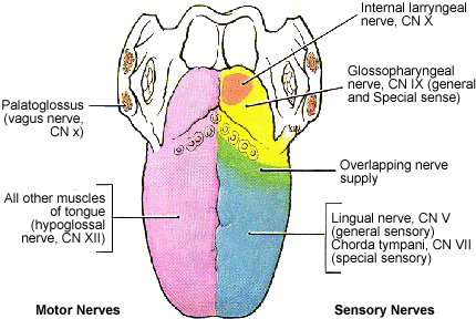
Nerve supply:
– Posterior 1/3 of tongue: General sensation and taste supplied by glossopharyngeal nerve (CN IX).
– Anterior 2/3 of tongue:
› General sensation: Lingual nerve (branch of mandibular division of CN V3)
› Taste: Chorda tympani nerve (branch of facial nerve CN VII)
– Hypoglossal nerve (CN XII): Provides motor innervation to all intrinsic and extrinsic tongue muscles except palatoglossus.
Extrinsic muscles:
– Styloglossus: Retracts and elevates tongue, CN XII.
– Hyoglossus: Depresses and retracts tongue, CN XII.
– Genioglossus: Protrudes and depresses tongue, CN XII.
– Palatoglossus: Elevates posterior tongue, CN X (vagus nerve).
Muscles retracting tongue:
– Styloglossus: Pulls tongue posteriorly and superiorly.
– Hyoglossus: Pulls tongue posteroinferiorly.
The tongue receives sensory innervation from CN IX (posterior 1/3) and CN V3/VII (anterior 2/3) which provides general sensation and taste reception. Motor function and mobility are provided by CN XII which supplies most extrinsic tongue muscles. The palatoglossus is unique in receiving motor supply from CN X.
The styloglossus and hyoglossus muscles specifically act to retract and pull the tongue posteriorly. Along with other muscles, they allow for the extensive mobility and function of the tongue in speech, swallowing and gustation.
Q. Facial N. : Inra-cranial Course? Branches?
Origin: The facial nerve (CN VII) originates from the facial nucleus in the pons. It emerges between the pons and medulla, passes through the internal acoustic meatus (IAM) and then courses through the facial canal (aqueduct) in the petrous temporal bone.
Intra-cranial course:
1. Emerges between pons and medulla
2. Enters internal acoustic meatus (IAM)
3. Travels through facial canal in petrous temporal bone
4. Exits skull through stylomastoid foramen.
Branches: The main branches of the facial nerve in the face are:
1. Temporal branches: Supply frontal belly of occipitofrontalis and auricular muscles.
2. Zygomatic branches: Supply orbicularis oculi and zygomatic muscles.
3. Buccal branches: Supply buccinator and lip muscles.
4. Marginal mandibular branch: Supplies depressor anguli oris, depressor labii inferioris, and lower lip.
5. Cervical branch: Supplies platysma muscle in neck.
The facial nerve carries motor signals from the facial nucleus to the muscles of facial expression and a small glandular component (lacrimal gland). Damage to the facial nerve within the facial canal or after its exit from the stylomastoid foramen can lead to ipsilateral facial paralysis (Bell’s palsy) with loss of expression and lacrimation.
Recovery depends on the location of injury to the nerve. Proximal injuries near the nucleus have the lowest recovery, while distal injuries tend to recover spontaneously and with rehabilitation.
Q. Pterion forming bones? Significance?
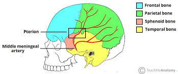
The pterion is formed by the articulation of four bones in the skull:
1. Frontal bone
2. Parietal bone
3. Sphenoid bone (greater wing)
4. Temporal bone (squamous part)
The clinical significance of the pterion is:
1. The middle meningeal artery passes closely deep to the pterion as it travels between the temporalis and temporoparietal fascia to enter the skull through the foramen spinosum.
2. Injury or fracture through the pterion can lacerate or rupture the middle meningeal artery, causing an epidural hematoma. This is a surgical emergency as the buildup of blood can compress the brain and cause herniation.
3. The pterion is a relatively weak point in the skull and fractures often pass through its region. Due to the proximity of middle meningeal artery, most pterion fractures require surgical management to prevent or evacuate an epidural hematoma.
4. Burr hole placement for emergency drainage of epidural or subdural hematomas is often placed over the pterion for direct access to the middle meningeal artery epidural space.
5. The frontal and temporal lobes of the cerebrum lie deep to the pterion, making it a commonly used surgical entry point for lobectomies or to gain intracranial access.
In summary, the pterion is an important clinical landmark formed by articulations of four skull bones. Its significance lies in the proximity of the middle meningeal artery deep to it, making pterion injuries or fractures a risk factor for epidural hematoma. It is also used as a surgical landmark and access point for burr hole and craniotomy placement.
Q. ID : Ethmoidal sinus / Coronal suture ( skull x-ray)?
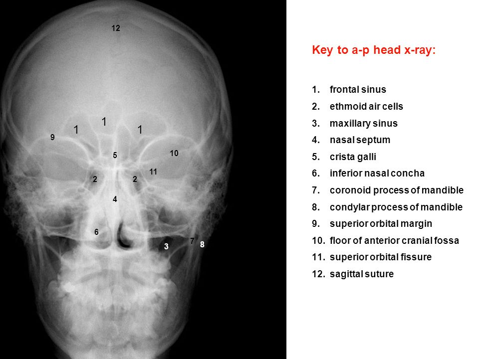
Ethmoidal sinuses:
– The ethmoidal sinuses occupy the ethmoid bone, located between the nasal cavity and the orbits. They appear as obliquely oriented air-filled spaces on skull X-ray.
– On the lateral view, the anterior and posterior ethmoidal sinuses are visible as two clusters of air cells separated by the basal lamella of the ethmoid bone.
– On the PA view, the ethmoidal sinuses form an irregular honeycomb pattern of air cells in the midline, superior to the nasal cavity and inferior to the frontal sinuses.
– The ethmoidal sinuses drain into the middle meatus of the nasal cavity via the middle ethmoidal canal. Their imaging appearance varies depending on the degree of pneumatization.
Coronal suture:
– The coronal suture separates the frontal bone anteriorly from the two parietal bones posteriorly.
– On the lateral skull X-ray, the coronal suture is seen as a shallow zig-zag line extending from the pterion superiorly to the temporal bone inferiorly.
– On the AP or PA view, the coronal suture extends from the greater wing of the sphenoid medially outwards beneath the temporal fascia, separating the frontal squama anteriorly from the parietal bones posteriorly.
– The coronal suture usually closes by adulthood but remains patent to some degree in most individuals. Premature closure of the coronal suture (craniosynostosis) causes characteristic skull deformities.
– The coronal suture serves as an important anatomical landmark to distinguish frontal from parietal bones on skull X-ray.
In summary, the ethmoidal sinuses appear as honeycomb patterns of air cells in the midline, while the coronal suture is seen as a shallow zig-zag line separating the frontal bone from parietal bones. Their appearances on X-ray can help distinguish regions and landmarks in the anterior and middle cranial fossae.
Q. Carotid canal contents ?
The carotid canal contains several important structures:
1. Internal carotid artery (ICA): Provides arterial blood supply to the brain and orbits. Ascends through the carotid canal, enters the cranium, and divides into the anterior and middle cerebral arteries.
2. Internal carotid nerve plexus: A network of postganglionic sympathetic fibers that surround the ICA.
3. Deep petrosal nerve: Provides sympathetic innervation to the sphenopalatine ganglion from the carotid plexus. Exits the skull through the foramen lacerum.
4. Emissary veins: Valveless veins that connect the cavernous sinus to veins external to the skull, providing a potential route for infection spread. Examples include the emissary sphenoidal and parietal veins which traverse the carotid canal.
5. Caroticotympanic nerves: Provide sensory innervation to the middle ear, auditory tube and tympanic membrane. Branch from the glossopharyngeal nerve and the carotid plexus within the canal.
The carotid canal is found within the petrous temporal bone and transmits the internal carotid artery and associated sympathetic nerves into the cranium to supply the brain and orbit. It also allows for connections between intra-cranial and extra-cranial venous systems via the emissary veins – a potential weakness that can allow the spread of infection.
The structures transmitting through the carotid canal include:
ICA – Provides arterial blood supply to brain and orbits.
Carotid nerve plexus – Sympathetic innervation.
Deep petrosal n. – To sphenopalatine ganglion.
Emissary veins – Connect cavernous sinus to external veins.
Caroticotympanic nn. – Sensory to middle ear, auditory tube and eardrum.